WHAT ARE EXOSOMES?
Granules approximately 30 to 200 nm in diameter encapsulated in a lipid bilayer that are secreted by the cells that make up the living body, from unicellular organisms to higher organisms.In humans, exosomes are found in various body fluids including breast milk, saliva, and tears as well as the blood and urine.In 2007, Lotvall et al. discovered that nucleic acids called microRNA are contained in exosomes. In 2010, Kosaka et al. of the National Cancer Center Japan demonstrated that this microRNA is incorporated and functions inside the cells that receive the exosomes.
As exosomes secreted from pathological tissues such as cancer cells contain disease-related molecules, they can serve as targets for drug discovery and diagnostic drugs.
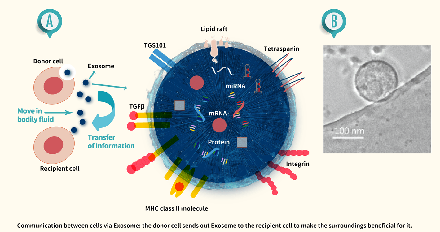
A.Structure of exosomes: The vesicle membrane consists of a phospholipid bilayer containing proteins such as tetraspanin. Exosomes contain proteins and nucleic acids (mRNA, miRNA). Modified from "Takahiro Ochiya, and Yusuke Yoshioka, Eds.
*Exosomes that Change Medicine: From Biological Functions to Disease Mechanisms and Clinical Applications. Kagaku Dojin, 2018, p. 8, Figure 2.1" (modified and reproduced with the permission of the copyright holder)
B.Morphological observation of exosomes under phase-contrast electron microscopy. Exosomes secreted by adipose-derived mesenchymal stem cells were collected by ultracentrifugation and observed under a phase-contrast electron microscope.
*Scale bar: 100 nm (provided by Yusuke Yoshioka)
EXOSCREEN: A SIMPLE AND HIGHLY SENSITIVE EXOSOME ASSAY
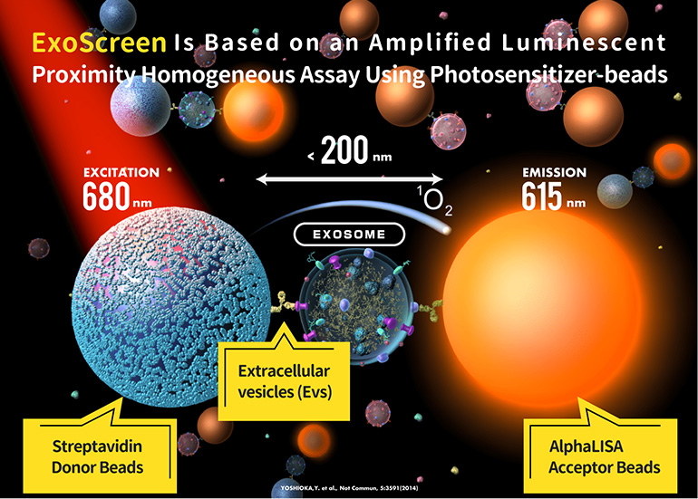
ExoScreen uses the principles of the measurement system AlphaLISA (PerkinElmer) and applies them to exosome measurement. This system uses the fact that the maximum range of singlet oxygen generated upon excitation at 680nm is 200nm, which roughly corresponds to the maximum diameter of exosomes. ExoScreen first binds the antibodies for CD9, an antigen found on the surface of all exosomes, to the acceptor beads. The antibodies for the cancer specific antigens thought to be found on the surface of exosomes secreted by cancer cells are then biotinylated and bound to donor beads with streptavidin. By detecting the light emitted when these two types of beads approach exosomes with both CD9 and cancer-specific antigens on the surface, ExoScreen quantifies the cancer-derived exosomes. As measurements with ExoScreen can be conducted simply by adding a blood sample to the reaction solution containing the beads, the test is extremely simple compared to the commonly used ELISA method. Theoria Science has filed a patent application for ExoScreen (International Patent Publication No.: WO2013/094307A1) and is also simultaneously registering the “ExoScreen” trademark.
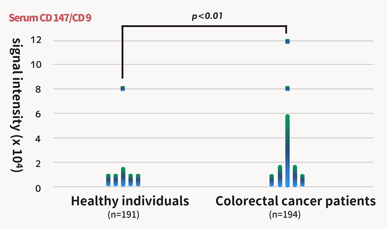
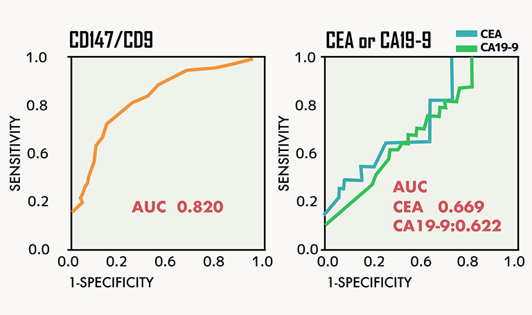
SERUM DERIVED FROM COLORECTAL CANCER PATIENTS AND SERUM DERIVED FROM HEALTHY INDIVIDUALS MEASURED USING EXOSCREEN
Upon measuring the signals derived from exosomes that express CD9 and CD147 in serum from colorectal cancer patients (194 subjects) and serum from healthy individuals (191 subjects), ExoScreen was shown to have a higher AUC, an indicator of diagnostic performance, compared with conventional blood tests (tumor marker CEA and CA 19-9). (AUC of the conventional method was approximately 0.65. AUC of ExoScreen was 0.82.) This is thought to enable the detection of early-stage cancer that could not be detected with the conventional blood tests.
*The higher the number for area under the curve (AUC), the higher the diagnostic performance.
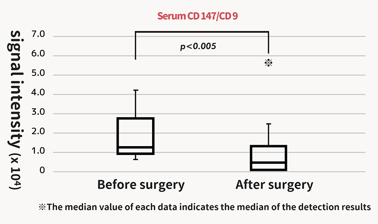
CHANGES IN SIGNAL INTENSITY BEFORE AND AFTER SURGERY
Changes in signals of exosomes that express CD9 and CD147 were examined before and after surgery using serum from 15 patients who underwent resection of stage I and II colorectal cancer. Tests using signals of exosomes expressing CD9 and CD 147 were found to be exceptional follow-up tests after surgery or drug administration.